Renal osteodystrophy and osteoporosis are not competing diagnoses. They are converging expressions of compromised skeletal health — and the patient pays for our therapeutic nihilism in fractures and cardiovascular events.
Coexistence is the rule, not the exception. The historical KDIGO separation of renal osteodystrophy from osteoporosis made them seem mutually exclusive — and the CKD patient pays the price in fractures.
The aging population delivers the typical patient as both a CKD case and an osteoporosis case. Declining eGFR routinely arrives on a skeleton already remodelled by menopausal bone loss, glucocorticoid exposure, smoking, immobility, and senescent low bone mass.1,9 CKD-MBD then develops on top of that pre-existing osteoporosis, not as its replacement.
Hip-fracture risk rises continuously with falling eGFR, with post-fracture mortality substantially higher than in matched controls.4,21,28 The premature-aging effect is striking: patients younger than 45 with CKD fracture at rates resembling those over 65 with normal renal function.22
Across large registries, nephrologists initiated denosumab in ~2.3% and oral bisphosphonates in ~2.6% of dialysis patients — a population whose absolute fracture risk vastly exceeds the postmenopausal women in whom these drugs are routinely prescribed.3,5 The bone specialist defers to the nephrologist; the nephrologist defers to the bone specialist; the patient fractures.
The deliverable of this guide is a sequenced, individualized framework that lets a nephrologist, internist, or endocrinologist make a defensible fracture-risk decision in CKD G1–G5D — without waiting for the perfect RCT that may never arrive.
Name the dominant lesion before treating. Turnover status reframes whether the first move is CKD-MBD control, antiresorptive, or anabolic.
Histologic ROD subtypes coexist with osteoporosis in any combination — they do not exclude it:
Under light microscopy, adynamic bone disease and low-turnover primary osteoporosis can be indistinguishable; osteoporosis has no formal histologic definition.24 Differentiation depends on clinical context (CKD stage, prior antiresorptive exposure, PTH trajectory, biochemistry). Do not start a potent antiresorptive in a patient with biochemical low turnover and an "osteoporotic" DXA without resolving this.
The 2023 Madrid Controversies Conference and subsequent EUROD / KDIGO output recentre the conversation on the skeleton, not the lab panel. "CKD-associated osteoporosis" reframes the patient as an osteoporosis patient whose treatment options are shaped — not replaced — by their CKD.7,9,10 This is the conceptual key that unlocks the therapeutic nihilism described in §1.
Drift to correct: a low T-score is consistent with — not synonymous with — osteoporosis. Other low-BMD states (osteomalacia, mixed ROD, secondary causes) are pathophysiologically distinct and respond differently to drug class.25,26
FGF-23 rises first. Klotho falls. PTH hyporesponsiveness is variable. Uremic toxins, inflammation, oxidative stress, and a calcification-paradox bone–vascular axis finish the job.
Interpret PTH against bone phenotype (turnover markers ± biopsy), not against a number on a guideline. A PTH of 300 pg/mL in one dialysis patient may indicate inadequately suppressed disease; in another, it may represent appropriate post-correction turnover. The 2025 KDIGO Controversies output explicitly endorses this individualization.9,12
Beyond classic risk factors, the uremic milieu actively degrades bone quality: uremic toxins, chronic low-grade inflammation, oxidative stress, gut dysbiosis, altered immunity, and accumulation of advanced glycation end-products.13,14 These drivers explain why a CKD patient with a "normal" BMD still fractures at an elevated rate — and why bone-strength rather than bone-density should anchor the diagnosis.
Patients with CKD lose mineral from the skeleton while gaining it in vessels — an inverse correlation between BMD and vascular calcification (VC) reproduced across cohorts.10,16 Proposed mechanistic links:
CKD is, in this sense, a model of accelerated aging: the same drivers that age the skeleton age the artery.13 This is why every treatment decision in §6 must be weighed on both ledgers.
Phosphate control, inflammation suppression, and oxidative-stress reduction may benefit bone and vasculature simultaneously. Whether bone-directed therapy modifies hard CV endpoints remains an open question — explicitly stated as a research gap in the source symposium.
Establish turnover before choosing the drug class. Most decisions can be made non-invasively — biopsy is reserved for the patient who refuses to fit the algorithm.
The CKD-associated osteoporosis panel goes beyond a DXA's biochemistry minimum:
| Domain | Test | Primary CKD use |
|---|---|---|
| Mineral chemistry | Calcium (albumin-corrected or ionised), phosphate | Stage-stratified targets per KDIGO 2017 / 2025 update.9,23 |
| PTH axis | Intact PTH (iPTH) | Trend, not single value — interpret against bone phenotype. |
| Vitamin D | 25(OH)D; selective 1,25(OH)₂D | Replace nutritional D before chasing activated forms.9 |
| Phosphaturic axis | FGF-23 (research / select) | Not routine; useful in hypophosphatemic outliers. |
| Bone formation | BSAP (bone-specific alkaline phosphatase), P1NP | Not renally cleared — reliable surrogates of turnover.27 |
| Bone resorption | TRAP-5b | Renal-resistant resorption marker. |
| Markers to avoid | β-CTX, NTX, P1NP (intact-form considerations) | Renally cleared — falsely high in advanced CKD; do not interpret as elevated turnover. |
Use a formation marker (BSAP or P1NP) together with a resorption marker (TRAP-5b). Concordantly high values support high turnover; concordantly low values support low-turnover / adynamic disease. Discordant values mean either mixed disease or assay/interference — a useful flag to obtain biopsy.
Tetracycline-labeled iliac-crest biopsy remains the reference standard when turnover or mineralization cannot be resolved non-invasively.9 Practical limits — invasiveness, labor, scarce histomorphometry labs, high interobserver variability, lack of normative data — are increasingly mitigated by AI-assisted quantification and TMV semiquantitative reads.
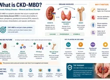
Bone and vessel share a single uremic biology. Every fracture-prevention decision must be weighed against the vascular ledger.
Arterial medial calcification and valvular calcification are highly prevalent in CKD G3–G5D, progress faster than in matched non-CKD controls, and are independently associated with cardiovascular and all-cause mortality.13,15,16 Downstream clinical sequelae:
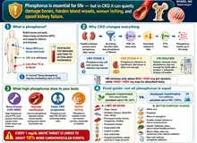
Hyperphosphatemia, elevated PTH, inflammation, oxidative stress, and uremic toxins drive osteoblast-like transdifferentiation of vascular smooth-muscle cells, with deposition of bone-matrix proteins in the vessel wall.15 The same milieu that suppresses bone formation in the skeleton ectopically promotes mineralization in the artery.
It is not a paradox. It is the predictable end-state of a single uremic biology expressed in two compartments. The shared-soil framing dictates the therapeutic posture: prioritise phosphate / inflammation / oxidative-stress control as a dual-benefit lever — and treat every bone-directed agent as a vascular decision too.
VC tracks with low BMD in primary osteoporosis (postmenopausal women, older men) as well — confirming a shared-soil phenomenon that CKD magnifies rather than invents.10,16
The §6 master algorithm requires an explicit weighing of vascular risk for each agent:
Organizing principle — CKD-MBD first, then bone-targeting. Correct mineral metabolism and suppress excess turnover before layering antiresorptive therapy. This sequence prevents most denosumab-in-dialysis catastrophes.
| Agent | eGFR window | Principal risk in CKD | Monitoring & sequence notes |
|---|---|---|---|
| Oral bisphosphonates (alendronate, risedronate) | Generally not initiated below eGFR ≈30–35 mL/min (FDA labelling; UK/EU guidance, verified 2026) | Prolonged skeletal retention; may worsen low-turnover/adynamic bone.19 | Individualize below threshold only with documented turnover. Stop after 3–5 yr with a drug-holiday plan. |
| IV zoledronate | Avoid < ~35 mL/min; risk of acute kidney injury and prolonged hypocalcemia in dialysis | AKI on infusion; hypocalcemia. | Not first-line in advanced CKD. |
| Denosumab (RANKL inhibitor) | Not renally cleared — effective in early CKD; FDA boxed warning for severe hypocalcemia in advanced CKD / dialysis (Jan 2024)3 | 41.1% severe hypocalcemia in dialysis women on denosumab vs 2.0% on oral bisphosphonates (Bird et al., JAMA 2024).3 Rebound vertebral-fracture risk on discontinuation. | Mandatory: correct CKD-MBD first; aggressive Ca + active vitamin D cover; Ca check days 3–10 post-dose; never stop without a follow-on antiresorptive. |
| Raloxifene (SERM) | Selected post-menopausal women on dialysis (observational) | VTE risk; modest BMD effect. | Second-line in HD women; document rationale. |
Do not initiate denosumab in a dialysis patient until you have: (1) corrected CKD-MBD — phosphate controlled, PTH appropriately suppressed, 25(OH)D repleted; (2) ensured adequate Ca + active vitamin D cover for the 10-day post-dose window; (3) scheduled Ca monitoring at days 3, 7, and 10 after each injection; (4) documented a follow-on antiresorptive plan at the time of initiation, not at the time of discontinuation. The Bird et al. 41.1% severe-hypocalcemia rate is what happens when steps 1–3 are skipped.3
A hypothesis-generating preventive frontier — explicitly not standard of care, but conceptually positioned to address mineral, skeletal, and vascular outcomes simultaneously.
Intermittent (not chronically elevated) PTH early in CKD — administered as FGF-23 begins to rise — could:
Reassuring in mild–moderate renal impairment in the Fracture Prevention Trial (teriparatide), ACTIVE (abaloparatide), and Japanese post-marketing data.2 Human cardiovascular endpoints in CKD specifically are not yet available.
Present this concept as "what's on the horizon," not as off-label clinical use. It is a strong candidate for a future intervention trial precisely because it targets the FGF-23 / Klotho / PTH axis at the stage where modification is still possible — and because it could plausibly close the bone-and-vessel ledger in a single intervention.
The single biggest gain from "one umbrella" is closing the renal/non-renal referral gap so neither side defers.
This clinician reference links out to the matching patient-facing material. Surface these when counselling the patient:
Most of what we do in CKD-associated osteoporosis is individualized, off-label, and shared-decision. Saying so out loud is the most honest part of the consent conversation.
Internal Medicine · Nephrology · Nutrition · Philippines · PRC 0105184
Last reviewed:
Educational. Decision support for licensed clinicians. Individualize to the patient, the local CKD-MBD lab availability, and the bone-specialist referral pathway.